


Introduction
Renal physiology is complicated. Don’t worry too much if you don’t feel like you have a full grasp of it – most of us don’t! It seems to be something that only medical registrars and renal physicians truly understand, and for the rest of us it can often remain a mystery. Nonetheless, I’ve attempted to get a handle on the basics in this article.
The job of the kidney is to remove waste products from the blood, as well as to regulate blood volume and plasma osmolarity. In the process, this creates urine.
- Urine consists of mainly water, with some salts and urea
- The kidneys receives a large blood supply to do this – about 20% of blood volume pumped each minute will pass through the kidneys
- Also remember that some of this process of removal of waste products (excretion) is done by the liver, and excreted in the faeces
A nephron is the basic functional unit of the kidney. Nephrons are microscopic in size, and a healthy adult has about 1 million nephrons in each kidney. Nephrons perform three roles; filtration, reabsorption, and secretion.
Firstly, nephrons filter the blood, creating a fluid we sometimes call filtrate. In the healthy kidney this should only contain water and small molecules. It shouldn’t contain any blood cells or any protein, but it contains most of the rest of the ‘stuff’ in the blood – and as such it is chemically similar to serum (without the protein). Secondly, the kidneys reabsorb many of the useful chemical constituents of this filtrate. Thirdly there is a process of exchange that occurs and some components of the blood (e.g. the larger proteins and other large molecules) can be actively secreted. Only once the filtrate has passed through all these processes do we call it urine.
- Excretion refers to the removal of waste from the body. Thus, the processes of filtration, reabsorption and secretion produce the end result of excretion.
The nephron is made up of:
- The renal corpuscle
- This bit does the filtering
- It is made up of the glomerulus and Bowman’s capsule
- The glomerulus is a bundle of blood vessels, surrounded by a semi-permeable membrane, which allows some of the constituents of the blood to flow through
- Bowmans capsule collects this fluid and delivers it to the renal tubule
- The renal tubule
- This bit does the reabsorption
- It also receives some of the secreted products from the efferent arteriole
- There are different sections of the tubule that have different roles in the process – which we will discuss later on
Blood arrives via the afferent arteriole into the glomerulus. It is filtered. The filtrate flows on into the renal tubule, whilst the blood flows into the efferent arteriole. The filtrate then has some of its constituents reabsorbed, and then the tubule and the efferent article meet up again at the location of the distal convoluted tubule, so that larger molecules can be actively secreted from the efferent arteriole into the tubule.

There are two types of nephrons, based on differences in their structure. The nephrons themselves sit across the boundary between between the medulla and the cortex of the kidney. Those that mostly sit in the cortex, are called cordial nephrons, and those that mostly sit in the medulla are called juxtamedually nephrons.
- 85% of nephrons are cortical nephrons and 15% are juxtamedullary nephrons.
- Juxtamedually nephrons are typically longer with a larger loop of Henle.

Filtration
There is a visceral epithelium that covers the glomerular capillaries inside Bowman’s capsule. This is just one cell thick, and it is made up of cells called podocytes. These podocytes have projections on their edge called pedicels. For stuff to pass out of the glomerular capillaries, it must be small enough to fit through the gaps between pedicels. These gaps are known as filtration slits.
The filtrate initially produced is pretty much the same as serum, but without the proteins.
In normal circumstances, no protein is able to leave and enter the filtrate. Any large molecules that have to be removed from the blood have to be actively removed further down the tubule.
The basement membrane around the glomerular capillaries is called the lamina densa and it is unusual in that cells of the lamina densa may attached to more than one capillary. This gives them greater control over capillary blood flow and capillary diameter.
The endothelium of the capillaries is fenestrated (has holes in it!). Together, this endothelium, the podocytes and the lamina densa make up the filtration membrane.
- Filtration is a passive process – the disadvantage of this being that you lose stuff you want to keep, such as glucose, vitamins, amino acids etc – and thus you have to get these back into the blood later on.
- The size of the ‘gaps’ in the filtration system varies. There are larger gaps in the capillaries than there are between the pedicels, and thus, placma proteins can pass out of the fenestrated capillaries, but cannot pass through the gaps between pedicels in normal renal functioning.
- BP in the glomerulus is atypically high for a capillary network. This is because the efferent arteriole has a smaller diameter than the afferent arteriole, and thus this keeps the pressure in the glomerulus high. Typically, pressure in the glomerulus are 50mmHg, as opposed to 35mmHg in other peripheral capillary beds.
- The pressure in the capsule opposes the pressure in the glomerulus – this pressure is about 15mmHG, and thus the net hydrostatic pressure is about 35mmHg.
- However, there is also colloid pressure due to the presence of proteins in the blood (and hopefully not in the filtrate!). This pressure is about 25mmHg. Thus, the overall pressure gradient is about 10mmHg, forcing solute in the direction of the tubules

Glomerular physiology. This file is taken from wikimedia commons and is licensed under the Creative Commons Attribution-Share Alike 3.0 Unported license.
Glomerular filtration rate
- The glomerular filtration rate (GFR) refers to the amount of fluid passing through all glomeruli in one minute.
- The GFR – normal GFR is about 125ml/minute.
- eGFR is a way of estimating GFR using an equation. The values required for calculation are serum creatinine, age, race, and sex.
- An eGFR is typically reported with blood results, alongside a blood tests for U+Es
- It is accurate in kidney disease, however, the method consistently underestimates the GRF of healthy people with a GFR of over 60ml/min.
- The GFR is kept pretty much constant in healthy individuals, despite normal fluctuations in blood pressure. For example a drop in blood pressure will cause:
- Dilation of the afferent arteriole
- Constriction of the efferent arteriole
- Dilation of the glomerular capillaries, and relaxation of the supporting cells.
- A rise in BP, will have the opposite effects. These effects are caused by baroreceptors in the wall of the afferent arteriole – if they are stretched (due to high bp), they will cause the arteriole to constrict – thus they oppose the changes.
- GFR is also regulated by hormonal changes – the renin-angiotensin system and natriuretic peptides
Creatinine clearance is another way we can measure GFR. We know that the concentration of creatinine in the blood is fairly stable. We also know that creatinine is freely filtered into the filtrate, as well as actively secreted into the filtrate in small amounts. Thus, we can use creatinine clearance as a measure of kidney function and to estimate GFR.
- Urine creatinine concentration is compared to plasma creatinine concentration. You measure how much creatinine a person excretes within a given time (e.g. 84 mg/hour). This works out at 1.4mg/minute. If we assume that the plasma concentration of creatinine is the same as the urine concentration, (which just happens to be 1.4mg/100ml), then we know that 100ml of plasma was filtered, and thus the GFR is 100ml/min.
Reabsorption and Secretion
Reabsorption can either be passive – by diffusion, or active, where substances are pumped against a concentration gradient from the tubule. Secretion refers to the active removal of molecules from the blood and into the tubular fluid.
Reabsorption occurs in the renal tubule. This can be divided into three main sections:
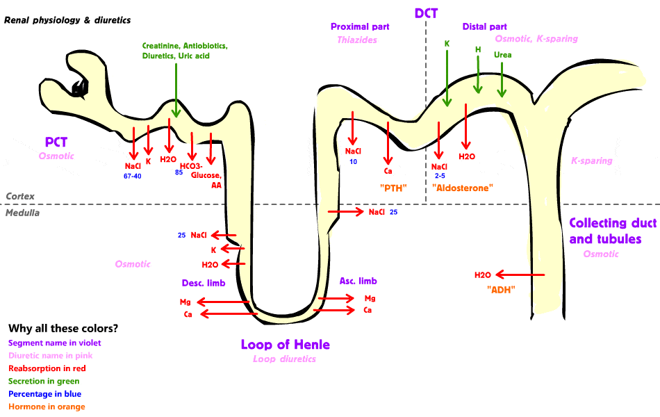
- Proximal convoluted tubule – PCT
- About 70% of the processes of reabsorption occurs here
- It also actively secreted large molecules – typically strong acids and bases – and as such this is where most drugs are secreted by the kidney
- The loop of Henle
- Causes concentration of the filtrate, by actively reabsorbing ions – such madly sodium, but also magnesium, potassium and calcium
- Water is then passively reabsorbed in response to this concentration gradient change
- The Distal Convoluted Tubule
- Its main job is secretion of ions into the filtrate – particularly potassium
- It can also reabsorb calcium
Proximal Convoluted Tubule
The PCT (proximal convoluted tubule) is primarily involved in reabsorption – it has a cuboidal epithelium with many microvilli. This is the main place where organic molecules are reabsorbed. Reabsorption of ions and water also occurs here, and the cells do have the ability to secrete, although this is not their main function. Anything that is reabsorbed here is then secreted into the peritubular space (extracellular fluid) where it will be taken up by the peritubular capillaries.
- 70% of reabsoprtion occurs here. Sodium is very important in may PCT processes.The things that are reabsorbed are:
- Organic stuff – amino acids and what not are all removed by facilitated diffusion and co-transport. For example, glucose is taken up by co-transport with sodium.
- Active removal of ions – including sodium, potassium, bicarbonate, magnesium, phosphate and sulphate. Sodium is exchanged for hydrogen. The hydrogen then combines with bicarbonate in the filtrate, and this reacts to turn into water and carbon dioxide. The carbon dioxide is taken up by the PCT cell, where the previous reaction is reversed, releasing bicarbonate and hydrogen!. The bicarbonate is removed into the blood, whilst the hydrogen starts its loop cycles again.
- The ion pumps in this region can be directly affected by hormones (e.g. angiotensin II stimulates sodium reabsoprtion in the PCT).
- Water reabsoprtion – due to osmotic changes due to active ion reabsoprtion.
- Passive removal of ions – they follow the water
- At the base of the PCT cells sodium ions are actively pumped out by a sodium potassium pump. This helps drive many of the process of PCT re-absorption, such as glucose uptake, and bicarbonate re-absorption. At the filtrate side of the cell, sodium ions are attracted to the cell by both their concentration gradient, and a negative charge of the cell created by the sodium potassium pump. Sodium ions can then enter by passive diffuse, co-transport (with organic molecules) or counter transport (with H+, allowing the reabsoprtion of bicarbonate ions). Further along the PCT, concentrations of organic molecules is low, and thus not much sodium can be absorbed by con-transport. So in these instances, sodium is absorbed by co-transport with chloride.
- Potassium will passively diffuse out of the filtrate. However, the PCT also has an ability to secrete potassium if levels are very high.
- Secretion by the PCT is generally for strong acids and strong bases – this is generally secretion of DRUGS!.
Loop of Henle
In the loop of Henle, the ‘thick’ and ‘thin’ segments refer to the size of the cells of this region, and NOT to the diameter of the tubule – this remains roughly the same!
- The thick descending limb has a similar role to the PCT and basically pumps out ions and solutes into the peritubular fluid. This creates a high concentration of solutes in the peritubular fluid, and thus in the peritubular capillaries.
- The thin descending limb is only permeable to water, and thus water will passively cross its membrane owing to the high concentration of solutes in the peritubular fluid, created by the PCT and thick descending limb.
- The loop of Henle concentrates the tubular fluid. It will actively pump out lots of sodium and chloride from the tubular fluid.
- In the loop of Henle a phenomenon known as countercurrent multiplication occurs. This basically is the term for the concentration of the urine. The thick parts of the loop pump out ions, and are pretty much impermeable to everything else. The thin part of the loop is permeable to water but not to anything else. Basically the thick part is the ascending part, and the thin part is the descending part. The thick part pumps out lots of ions, and this creates an osmotic gradient in the peritubular region near the descending limb. This then draws water out of the descending limb (before it reaches the thick ascending limb). This is the process of countercurrent multiplication.
- The pump that pumps out ions is the sodium/potassium/chloride pump. It pumps out 1 sodium, 1 potassium and 2 chloride ions, into the loop of Henle cells. However, the potassium ions later diffuse back into the tubular fluid, and thus the net result of this transport is that it removes 1 sodium and 2 chloride ions from the tubular fluid. Potassium ions are also exchanged for sodium and chloride at the base of the cell, but remember that the overall movement of potassium ions in this region is nil.
- Juxtamedually nephrons have a longer loop of Henle, and thus they concentrate the urine more.
- The vasa recta is a system of capillaries that is found surrounding the loop of Henle. These capillaries will exchange water and ions with the extracellular space around the loop of Henle. The capillaries will lose water and gain ions until equilibrium is reached. Water is drawn out of the capillaries as it is also drawn out of the descending loop of Henle. At the bottom of the loop of Henle this creates a capillary blood that is very thick. As the capillary network then travels back up the ascending loop of Henle it absorbs water from the interstitia. This water is drawn from the collecting ducts! – we have just seen how the ascending limb of Henle pumps out lots of ions, thus creating a hypotonic solution. This solution then travels down the collecting duct, alongside the loop of Henle, next to the concentrated blood of the vasa recta, and thus water moves from the collecting system into the capillaries of the vasa recta. This ensures as little urine as possible is lost.
- This process is controlled by ADH (vasopressin). This is a peptide hormone that is synthesised in the hypothalamus and secreted by the posterior pituitary. ADH essentially increases the permeability of the collecting ducts to water, thus allowing more water to be drawn out by the vasa recta, and thus creating a more concentrated urine.
Distal Convoluted Tubule
The DCT (distal convoluted tubule) – this has a thinner diameter than the PCT. It actually heads back towards the glomerulus, and passes between efferent and afferent arterioles, before doing a U-turn and heading back the way it came from. It actively secretes ions, selectively secretes sodium and calcium, and allows the reabsorption of water. Its main job it the further removal of sodium and chloride from the tubular fluid. Aldosterone will increase the rate of this by increasing the synthesis and mobilization of the sodium channels. Essentially, by the time tubular fluid gets to the collecting ducts, there is only water, urea, creatinine, potassium, hydrogen and maybe some urobillogens / stercobillogens left in it! There isn’t much!
- Retention of sodium (i.e. in the presence of aldosterone) is associated with loss of potassium.
- The DCT is also the place where calcium is reabsorbed – a process that is controlled by parathyroid hormone and calcitriol.
- Hydrogen ions are also secreted in this region in some pretty complicated ways! This is important in controlling blood pH.
The Collecting System
The collecting system – allows secretion of hydrogen and bicarbonate ions, and thus allows the control of blood pH. It also allows the reabsoprtion of bicarbonate, sodium and urea in varying amounts.
The juxtaglomerular apparatus refers to cells of the DCT in juxtaglomerular nephrons that are at the site of the efferent and afferent arterioles. The cells in this region are specialized, and referred to as the macula densa. Together with unusual cells of the afferent arteriole, the macular densa makes up the juxtaglomerular apparatus. These cells are endocrine in function, and secrete erothropoiten and renin.
The three most important waste products in urine are urea, creatinine and uric acid. Urea comes mainly from the breakdown of amino acids, creatinine is a waste product of creatinine phosphate produced by muscle contraction, and uric acid is a waste product from the recycling of RNA molecules. These products can only be excreted when dissolved in water – and thus excretion of them results in unavoidable water loss.
The kidneys can produce a solution that is 4x as concentrated as plasma
The renal threshold is the concentration at which a substance will no longer be able to be completely reabsorbed, and thus will begin to appear in the urine. For example, glucose has a renal threshold. Once concentration in the blood exceeds this threshold, then glucose will appear in the urine. As a substance approaches its renal threshold, its rate of removal from tubular fluid also increases because this is related to its concentration. Glucose renal threshold is about 10mmol/L. so when blood glucose levels are higher than this, the PCT cannot remove all the glucose from the filtrate, and glucose starts to appear in the urine.
The renal threshold for water soluble vitamins is particularly low, and thus if you take vitamin supplement, you are likely to just pee them all out!

Secretion or Excretion?
- Secretion refers to the process of active transfer of a molecule from one place to another – in the case of the kidney it refers to products being secreted into the tubule
- Excretion refers to the process of removal of waste products from the body. Thus the kidney is involved with excretion, and the whole process of filtration, reabsorption and secretion can be thought of collectively as excretion.
References
- Guyton, AC., Hall, JE. (2005). Textbook of Medical Physiology. Saunders
- Murtagh’s General Practice. 6th Ed. (2015) John Murtagh, Jill Rosenblatt
- Oxford Handbook of General Practice. 3rd Ed. (2010) Simon, C., Everitt, H., van Drop, F.
- Beers, MH., Porter RS., Jones, TV., Kaplan JL., Berkwits, M. The Merck Manual of Diagnosis and Therapy