Contents
Introduction
This article looks at adrenal physiology and explains the production and effects of adrenal products. For more information on mechanisms of blood pressure regulation, secretion of ions, and other effects related to the action of mineralocorticoids, please see the control of renal function.
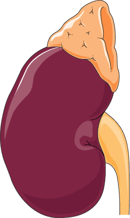
- The adrenal glands are also sometimes known as suprarenal glands
- They sit on top of the kidneys. They weight about 4g, and have a medulla and a cortex, like the kidneys themselves.
- The medulla – is directly connected to the sympathetic nervous system, and will secrete adrenaline and noradrenaline in response to sympathetic stimulation.
- These two hormones cause almost the exact same effects on body tissues as direct sympathetic stimulation itself does.
- The cortex secretes an entirely different type of hormone – corticosteroids. The corticosteroids are all synthesised from cholesterol, and have similar chemical formulas.
- There are three types of corticosteroid:
- Mineralcorticoids – e.g. aldosterone – these are so called because they effect the ‘minerals’ (electrolytes) of the blood. They particularly effect sodium and potassium.
- Glucocorticoids – e.g. cortisol – so called due to their effects on glucose metabolism – however they also have important effects on protein and fat metabolism.
- Androgens – these are sex hormones that exhibit similar effects to testosterone. They are not particularly important, except in disease of the adrenal glands where their hypersecretion can result in masculization.

Anatomy
- The zona glomerulosa – this constitutes about 25% of the adrenal cortex, and it is where aldosterone is produced – this region contains the enzyme aldosterone synthase. The production of aldosterone is controlled by extracellular fluid concentrations of angiotensin II and potassium. Both these two chemicals will increase the synthesis of aldosterone. Prolonged stimulation of this zone can lead to its hypertrophy.

Cross section of adrenal gland - The zona fasciculata – this constitutes about 75% of the adrenal cortex, and secretes glucocorticoids as well as small amounts of androgens and oestrogens. The secretion of these hormones is largely controlled by the hypothalamic-pituitary axis – and the release of ACTH (adrenocorticotropic hormone).
- The zona reticularis – this is responsilbe for most of the androgen output of the adrenal galnd and it also secretes some oestrogens and glucocorticoids.
- The mechanisms of androgen secretion are not well understood in comparison with the mechanisms of mineralcorticoid and glucocorticoid secretion.
Corticosteroids
Often a ‘mineralocorticoid’ will also have some glucocorticoid activity, and vice-versa.
- ACTH will cause an increase in corticosteroid synthesis by both increasing the number of surface receptors for LDL’s and it will also increase the number of enzymes used to liberate cholesterol from endocytosed LDL’s.
- Note that stress also causes an increase in cortisol production.
- The rate limiting step in to production of adrenal hormones is the first step in cholesterol breakdown, which occurs in the mitochondria by the enzyme cholesterol desmolase.
- ACTH and angiotensin II will both increase the rate of this reaction.
- There are then a series of reactions in the mitochondria, and then later on the ER that lead to the production of hormones. Obviously, some of these reactions differ depending on which region of the cortex you are in.
The general effect of these binding globulins is that it helps buffer sudden changes in concentration of the corticosteroids, such as in brief periods of stress, or when ACTH is released. Binding proteins also ensure that there is a uniform distribution of corticosteroids to the peripheral tissues.
Diseases of the liver directly reduce the excretion of these products.
Mineralocorticoids
Many mineralocorticoids also have glucocorticoid activity!
Cortisol is particularly relevant in pathological instances – it has relatively little mineralocorticoid activity, but in excess this can become noticeable – however it will also cause significant glucocorticoid issues!
Effects of aldosterone
- Aldosterone causes increased absorption of sodium, and increased secretion of potassium by the renal tubules. Therefore, in the extracellular fluid, aldosterone causes an increase in sodium and a decrease in potassium levels.
- Conversely, a lack of aldosterone can result in high extracellular potassium levels and low sodium levels.
- Despite these effects, the actual amount of sodium retained is very small, and thus excess sodium retention is rarely an issue. However, the sodium retention causes secondary fluid retention and thus increases arterial pressure significantly. It also causes a sensation of thirst.
- Any excess fluid and sodium will then be removed by normal kidney function – due to pressure diuresis. This method of maintaining normal fluid and salt levels despite high levels of aldosterone is known as the aldosterone escape. Once this level is reached, the level amount of salt and water gain by the body is zero. However, by this stage, the individual will be in a state of hypertension.
- Excess aldosterone will cause hypokalaemia which will lead to muscle weakness as a result of altered cell permeability. It also leads to alkalosis; because aldosterone causes retention of sodium – and this occurs by two mechanisms. The first (already discussed) is where potassium is exchanged for sodium in the renal tubules, but another mechanism involves the exchange of sodium for hydrogen, thus resulting in mild alkalosis. Lack of aldosterone can lead to cardiac failure as a result of high potassium levels.
- Aldosterone also has effect on the GIt and on sweat glands.
- Sweating – normally, we secrete sodium chloride, potassium and bicarbonate in our sweat. The bicarbonate and potassium tend to be subsequently lost, but the sodium chloride can be re-absorbed along the sweat gland. In the presence of aldosterone, this process is enhanced.
- GIt – aldosterone enhances the absorption of sodium from the diet. This effect mainly occurs in the colon. Without aldosterone, this sort of sodium absorption can be poor. This also means that fewer chloride ions and less water is absorbed, which can in turn cause diarrhoea, exaggerating the effect.
Mechanism of action
- Potassium ion concentration
- Renin-angiotensin system
Glucocorticoids
Mechanism
Metabolic actions
- Carbohydrates – Glucocorticoids cause a decrease in the utilization of circulating glucose (mechanism unknown – thought o involve modification of enzymes involved with glucose breakdown within a cell), and an increase in gluconeogenesis. This leads to a tendency for hyperglycaemia. There is also an increase in glucose storage, which is probably a result of increased secreted insulin as a response to the hyperglycaemia.
- Proteins – causes increased catabolism and decreased anabolism – i.e. they cause an overall increase in ‘metabolism’ – breakdown of products to release energy, but a decrease in ‘growth’. Overall there is an increase in protein breakdown, and a decrease in protein synthesis – which can lead to muscle ‘wasting’. However, there is an increase in synthesis of liver proteins. This effect is thought to result from an enhanced transport of amino acid into liver cells (whereas with most other cells, amino acid transport into cells in decreased – but catabolism carries on as normal, and thus over time, proteins are removed from peripheral cells).
- Fats –Initially it causes mobilisation of fat from adipose tissue into the blood, allowing their utilisation for energy needs. This effect probably results from reduced glucose transport into adipose cells, thus they think that glucose levels are low, and so they release fats.
- Later, it has an effect on lypolitic hormones, and causes a redistribution of fat, like that seen in Cushing’s syndrome (e.g. Moon face and buffalo hump). This peculiar type of obesity results from the deposition of fat on the torso and around the head.
- Electrolytes – glucocorticoids tend to reduce the amount of calcium in the body by reducing its uptake from the GIt, and increasing its excretion by the kidneys. This can induce osteoporosis. Glucocorticoids are also likely to cause sodium retention and potassium loss.
Regulatory actions
Hypothalamus and anterior pituitary – causes a feedback effect resulting in reduced release of endogenous glucocorticoids
Cardiovascular system – reduced vasodilation and decreased fluid exudation (oozing)
Musculoskeletal system – decreased osteoblast, and decreased osteoclast activity
Inflammation and immunity
- Acute inflammation – decreased influx and activity of leukocytes
- Chronic inflammation – decreased activity of mononuclear cells, decreased angiogenesis (development of new blood vessels)
- Lymphoid tissues – decreased action of B and T cells, and decreased release of inflammatory mediators by T cells.
- Decreased production of cytokines
- Decreased expression of COX-2 and thus decreased prostaglandin synthesis
- Decreased generation of nitric oxide
- Decreased histamine release from basophils
- Decreased production of IgG
- Decreased complement components in the blood
- Increased anti-inflammatory factors such as IL-10 and annexin-1
- Overall, this results in decreased immune response – both to acquired auto-immune problems, but also to the protective role of the immune system.
Cortisol and stress
- Trauma
- Infection
- Extreme temperature
- Surgery
- Almost any debilitating disease!
- Environmental / social factors – feeling ‘stressed’!
However, it is not always certain why cortisol and its effects are useful in stressful situations. The most obvious thing is that glucose is made available for utilisation, although its actual utilisation is slowed and impeded. Also it is thought that the proteins released in catabolism can, in the very short term, be used by cells to create proteins that are essential to life (such as in the case of trauma). This is possible due to the fact that the most important proteins in a cell are affected last by cortisol – i.e. the ones you need to least are the ones that are broken down first.
Anti-inflammatory effects
- Stabilisation of lysosomal membranes – this makes it much more difficult for membranes of lysosomes to rupture, and thus the inflammatory proteins often released in the first stages of inflammation are much less likely to be released.
- Decreased capillary permeability – this is probably secondary to the first effect.
- Decreased migration and activity of white blood cells – this is a result of reduced release of inflammatory proteins.
- General immune system suppression – especially that of T cells. This reduces inflammation, due to the inflammatory actions of T cells on affected areas.
- Reduction of fever – this is mainly a result of reduced secretion of IL-1 from white blood cells. The reduced temperature will also reduce the amount of vasodilation, and thus reduce oedema.
One final effect is that cortisol increases the number of RBC’s. Conversely a lack of cortisol can result in anaemia.
Regulation of secretion
Glucocorticoids as treatments
Unwanted effects
- Poor wound healing
- Peptic ulceration
- Cushing’s syndrome – which is basically a manifestation of all the metabolic and systemic effects described above.
- Diabetes – as a result of the hyperglycaemia
- Weakness and muscle wasting
- Stunted growth in children – particularly if the treatment is continued for more than 6 months – even if the dose is low.
- CNS effects – often the patient may experience euphoris, but it can also manifest as depression. In depressed patients, the depression may be due to a disruption of the circadian rhythm secretion of the steroids.
- Oral thrush (candidasis) often occurs when the drugs are taken orally, as a result of suppression of local inflammatory processes.
Pharmacokinetics
References
- Murtagh’s General Practice. 6th Ed. (2015) John Murtagh, Jill Rosenblatt
- Oxford Handbook of General Practice. 3rd Ed. (2010) Simon, C., Everitt, H., van Drop, F.
Read more about our sources


